
Les 16, 18 et 19 septembre 2026des examens au microscope des iPhone 18 Pro et 18 Pro Max ont montré une modification de la dalle OLED que la présentation du produit ne détaillait pas : une partie du système infrarouge de Face ID passe sous l’écran.
La Dynamic Island mesure 16 mm de long, contre 21 mm sur la génération précédente, pour une épaisseur proche de 6 mm. Apple associe cette évolution à une réduction de 25 % de la surface occupée par l’îlot.
Le changement ne vient pas d’une retouche graphique. Il concerne l’organisation des sous-pixels dans la partie supérieure gauche de la dalle, près de l’heure. Le microscope permet de voir la zone réservée au composant déplacé.
Le gain se paie ailleurs.
Face ID déménage, la pilule raccourcit
Sur les générations précédentes, la Dynamic Island regroupait trois éléments de Face ID : la caméra frontale, l’émetteur infrarouge et le récepteur infrarouge. Sur les iPhone 18 Pro, le récepteur quitte l’îlot et passe sous l’écran.
Le composant déplacé n’est donc pas l’appareil photo frontal. Le récepteur recueille les informations infrarouges utilisées par Face ID pour analyser le visage en trois dimensions, mais il les reçoit désormais à travers la dalle OLED.
La nouvelle architecture libère l’espace occupé par le récepteur dans l’îlot. Elle explique le passage de 21 à 16 mm en longueur, alors que l’épaisseur reste proche de 6 mm.
L’espace récupéré permet à Apple d’afficher une activité en direct supplémentaire à côté de la Dynamic Island. Les iPhone 18 Pro et 18 Pro Max peuvent ainsi en présenter trois simultanément, contre deux sur les autres modèles.
Sous l’écran, la couture des pixels
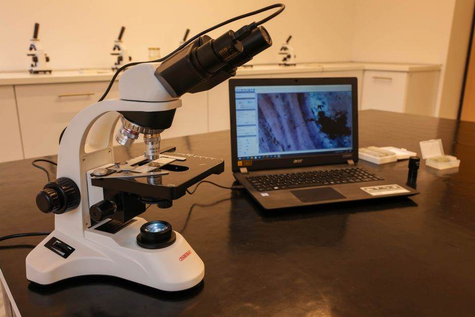
Un écran OLED classique repose sur une matrice de sous-pixels rouges, verts et bleus. Elle affiche l’image, mais laisse peu de passages libres à la lumière infrarouge. Le récepteur a donc besoin d’une zone où cette lumière peut atteindre le capteur.
À fort grossissement, la matrice reste régulière dans la majeure partie de l’écran, puis devient moins dense au niveau du récepteur. La densité de sous-pixels y apparaît environ deux fois plus faible.
Le motif observé ne correspond pas à un simple trou transparent. Les sous-pixels adoptent une disposition en chevrons qui laisse passer des lignes continues de lumière infrarouge. L’écran continue d’afficher une image à cet endroit, mais sa trame laisse davantage circuler le signal utilisé par Face ID.
Le récepteur passe sous la dalle. Les sous-pixels s’espacent dans cette zone. La Dynamic Island perd cinq millimètres.
Le microscope révèle cette couture.
Un rond visible, mais pas partout

À l’oeil nu, la zone modifiée reste difficile à distinguer. Il faut réunir un éclairage particulier et un angle de vision précis pour apercevoir la différence dans la partie supérieure gauche de l’écran, près de l’heure.
Dans certaines conditions, un petit rond peut apparaître à cet endroit. Il correspond à la zone de sous-pixels moins dense qui laisse passer les infrarouges vers le récepteur de Face ID.
Une photo prise en macro rend les écarts entre les sous-pixels beaucoup plus lisibles qu’à distance normale. Une zone visible sur ce type de cliché ne l’est donc pas forcément pendant l’utilisation. La trace peut apparaître dans certaines conditions sans rester constamment visible.
À distance normale, la zone est beaucoup moins facile à repérer. La dalle n’a toutefois pas la même densité de sous-pixels partout : la différence reste inscrite dans sa construction, même lorsqu’elle échappe au regard.
Le microscope ne teste pas le déverrouillage
Le microscope répond à une question d’intégration : où se trouve le récepteur et comment l’écran laisse-t-il passer le signal infrarouge? Il ne mesure ni la vitesse ni la fiabilité de la reconnaissance faciale.
L’évaluation de l’usage relève d’autres tests, notamment dans l’obscurité et lorsque l’angle du visage change. La structure de la dalle montre le compromis matériel, pas le comportement complet de Face ID.
Le récepteur infrarouge n’a pas les mêmes exigences qu’une caméra selfie : il doit recevoir un signal exploitable, et non produire une image de haute qualité pour l’utilisateur. Cette différence de fonction explique pourquoi la caméra frontale reste dans la Dynamic Island alors que le récepteur passe sous l’écran.
Les iPhone 18 Pro ne suppriment donc pas l’ouverture frontale. La caméra et l’émetteur restent dans l’îlot, tandis que le récepteur se cache sous une zone de pixels moins dense.
La Dynamic Island gagne cinq millimètres, au prix d’une zone de pixels moins dense.
Sources et références scientifiques
- Electron microscopy ascertains presence of centrioles in rectal epithelial cells of C. elegans L1 larvae – PMC. microPublication biology. DOI: 10.17912/micropub.biology.001624 · PMID: 40475707.
- Dammermann A, Müller-Reichert T, Pelletier L, Habermann B, Desai A, Oegema K. Centriole assembly requires both centriolar and pericentriolar material proteins. Dev Cell. 2004 Dec 1;7(6):815-829. doi: 10.1016/vcel.2004.10.015. [DOI] [PubMed] [Google Scholar]. doi:10.1016/j.devcel.2004.10.015.
- Delattre M, Leidel S, Wani K, Baumer K, Bamat J, Schnabel H, Feichtinger R, Schnabel R, Gönczy P. Centriolar SAS-5 is required for centrosome duplication in C. elegans. Nat Cell Biol. 2004 Jul 1;6(7):656-664. doi: 10.1038/ncb1146. [DOI] [PubMed] [Google Scholar]. doi:10.1038/ncb1146.
- Fırat-Karalar EN, Stearns T. The centriole duplication cycle. Philos Trans R Soc Lond B Biol Sci. 2014 Sep 5;369(1650) doi: 10.1098/rstb.2013.0460. [DOI] [PMC free article] [PubMed] [Google Scholar]. doi:10.1098/rstb.2013.0460.
- Kalbfuss N, Gönczy P. Extensive programmed centriole elimination unveiled in C. elegans embryos. Sci Adv. 2023 May 31;9(22):eadg8682-eadg8682. doi: 10.1126/sciadv.adg8682. [DOI] [PMC free article] [PubMed] [Google Scholar]. doi:10.1126/sciadv.adg8682.
- Kalbfuss N, Gönczy P. Towards understanding centriole elimination. Open Biol. 2023 Nov 15;13(11):230222-230222. doi: 10.1098/rsob.230222. [DOI] [PMC free article] [PubMed] [Google Scholar]. doi:10.1098/rsob.230222.
- Kalbfuss N, Berger A, Gönczy P. Mapping of centriolar proteins onto the post-embryonic lineage of C. elegans. Dev Biol. 2023 Jul 4;502:68-76. doi: 10.1016/j.ydbio.2023.07.001. [DOI] [PubMed] [Google Scholar]. doi:10.1016/j.ydbio.2023.07.001.
- Kemp CA, Kopish KR, Zipperlen P, Ahringer J, O'Connell KF. Centrosome maturation and duplication in C. elegans require the coiled-coil protein SPD-2. Dev Cell. 2004 Apr 1;6(4):511-523. doi: 10.1016/s1534-5807(04)00066-8. [DOI] [PubMed] [Google Scholar]. doi:10.1016/s1534-5807(04)00066-8.
- Kirkham M, Müller-Reichert T, Oegema K, Grill S, Hyman AA. SAS-4 is a C. elegans centriolar protein that controls centrosome size. Cell. 2003 Feb 21;112(4):575-587. doi: 10.1016/s0092-8674(03)00117-x. [DOI] [PubMed] [Google Scholar]. doi:10.1016/s0092-8674(03)00117-x.
- Leidel S, Gönczy P. SAS-4 is essential for centrosome duplication in C elegans and is recruited to daughter centrioles once per cell cycle. Dev Cell. 2003 Mar 1;4(3):431-439. doi: 10.1016/s1534-5807(03)00062-5. [DOI] [PubMed] [Google Scholar]. doi:10.1016/s1534-5807(03)00062-5.
- O'Connell KF, Caron C, Kopish KR, Hurd DD, Kemphues KJ, Li Y, White JG. The C. elegans zyg-1 gene encodes a regulator of centrosome duplication with distinct maternal and paternal roles in the embryo. Cell. 2001 May 18;105(4):547-558. doi: 10.1016/s0092-8674(01)00338-5. [DOI] [PubMed] [Google Scholar]. doi:10.1016/s0092-8674(01)00338-5.
- Pelletier L, Ozlü N, Hannak E, Cowan C, Habermann B, Ruer M, Müller-Reichert T, Hyman AA. The Caenorhabditis elegans centrosomal protein SPD-2 is required for both pericentriolar material recruitment and centriole duplication. Curr Biol. 2004 May 25;14(10):863-873. doi: 10.1016/j.cub.2004.04.012. [DOI] [PubMed] [Google Scholar]. doi:10.1016/j.cub.2004.04.012.
- Pierron M, Woglar A, Busso C, Jha K, Mikeladze-Dvali T, Croisier M, Gönczy P. Centriole elimination during Caenorhabditis elegans oogenesis initiates with loss of the central tube protein SAS-1. EMBO J. 2023 Nov 21;42(24):e115076-e115076. doi: 10.15252/embj.2023115076. [DOI] [PMC free article] [PubMed] [Google Scholar]. doi:10.15252/embj.2023115076.
- Ultrastructure Expansion Microscopy applied to C. elegans embryos – PMC. microPublication biology. DOI: 10.17912/micropub.biology.001033 · PMID: 38774216.
- Askjaer P, Galy V, Meister P. Modern tools to study nuclear pore complexes and nucleocytoplasmic transport in Caenorhabditis elegans. Methods Cell Biol. 2014;122:277-310. doi: 10.1016/B978-0-12-417160-2.00013-8. [DOI] [PubMed] [Google Scholar]. doi:10.1016/B978-0-12-417160-2.00013-8.
- Beck M, Hurt E. The nuclear pore complex: understanding its function through structural insight. Nat Rev Mol Cell Biol. 2016 Dec 21;18(2):73-89. doi: 10.1038/nrm.2016.147. [DOI] [PubMed] [Google Scholar]. doi:10.1038/nrm.2016.147.
- Cohen M, Tzur YB, Neufeld E, Feinstein N, Delannoy MR, Wilson KL, Gruenbaum Y. Transmission electron microscope studies of the nuclear envelope in Caenorhabditis elegans embryos. J Struct Biol. 2002 Dec 1;140(1-3):232-240. doi: 10.1016/s1047-8477(02)00516-6. [DOI] [PubMed] [Google Scholar]. doi:10.1016/s1047-8477(02)00516-6.
- Duerr JS. Immunohistochemistry. WormBook. 2006 Jun 19;:1-61. doi: 10.1895/wormbook.1.105.1. [DOI] [PMC free article] [PubMed] [Google Scholar]. doi:10.1895/wormbook.1.105.1.
- Ferreira J, Stear JH, Saumweber H. Nucleoporins NPP-10, NPP-13 and NPP-20 are required for HCP-4 nuclear import to establish correct centromere assembly. J Cell Sci. 2017 Jan 25;130(5):963-974. doi: 10.1242/jcs.196709. [DOI] [PubMed] [Google Scholar]. doi:10.1242/jcs.196709.
- Galy V, Mattaj IW, Askjaer P. Caenorhabditis elegans nucleoporins Nup93 and Nup205 determine the limit of nuclear pore complex size exclusion in vivo. Mol Biol Cell. 2003 Aug 22;14(12):5104-5115. doi: 10.1091/mbc.e03-04-0237. [DOI] [PMC free article] [PubMed] [Google Scholar]. doi:10.1091/mbc.e03-04-0237.
- Gambarotto D, Hamel V, Guichard P. Ultrastructure expansion microscopy (U-ExM). Methods Cell Biol. 2020 Jul 1;161:57-81. doi: 10.1016/bs.mcb.2020.05.006. [DOI] [PubMed] [Google Scholar]. doi:10.1016/bs.mcb.2020.05.006.
- Gambarotto D, Zwettler FU, Le Guennec M, Schmidt-Cernohorska M, Fortun D, Borgers S, Heine J, Schloetel JG, Reuss M, Unser M, Boyden ES, Sauer M, Hamel V, Guichard P. Imaging cellular ultrastructures using expansion microscopy (U-ExM). Nat Methods. 2018 Dec 17;16(1):71-74. doi: 10.1038/s41592-018-0238-1. [DOI] [PMC free article] [PubMed] [Google Scholar]. doi:10.1038/s41592-018-0238-1.
- Gönczy P, Schnabel H, Kaletta T, Amores AD, Hyman T, Schnabel R. Dissection of cell division processes in the one cell stage Caenorhabditis elegans embryo by mutational analysis. J Cell Biol. 1999 Mar 8;144(5):927-946. doi: 10.1083/jcb.144.5.927. [DOI] [PMC free article] [PubMed] [Google Scholar]. doi:10.1083/jcb.144.5.927.
- Hachet V, Busso C, Toya M, Sugimoto A, Askjaer P, Gönczy P. The nucleoporin Nup205/NPP-3 is lost near centrosomes at mitotic onset and can modulate the timing of this process in Caenorhabditis elegans embryos. Mol Biol Cell. 2012 Jun 27;23(16):3111-3121. doi: 10.1091/mbc.E12-03-0204. [DOI] [PMC free article] [PubMed] [Google Scholar]. doi:10.1091/mbc.E12-03-0204.
- Hamel V, Guichard P. Improving the resolution of fluorescence nanoscopy using post-expansion labeling microscopy. Methods Cell Biol. 2020 Aug 20;161:297-315. doi: 10.1016/bs.mcb.2020.07.002. [DOI] [PubMed] [Google Scholar]. doi:10.1016/bs.mcb.2020.07.002.
- Hawryluk-Gara LA, Shibuya EK, Wozniak RW. Vertebrate Nup53 interacts with the nuclear lamina and is required for the assembly of a Nup93-containing complex. Mol Biol Cell. 2005 Feb 9;16(5):2382-2394. doi: 10.1091/mbc.e04-10-0857. [DOI] [PMC free article] [PubMed] [Google Scholar]. doi:10.1091/mbc.e04-10-0857.
- 3-iodotyrosine fluorescent proteins for improved expansion microscopy – PMC. microPublication biology. DOI: 10.17912/micropub.biology.002234 · PMID: 42553565.
- Arganda-Carreras Ignacio, Kaynig Verena, Rueden Curtis, Eliceiri Kevin W, Schindelin Johannes, Cardona Albert, Sebastian Seung H. Trainable Weka Segmentation: a machine learning tool for microscopy pixel classification. Bioinformatics. 2017 Mar 30;33(15):2424-2426. doi: 10.1093/bioinformatics/btx180. [DOI] [PubMed] [Google Scholar]. doi:10.1093/bioinformatics/btx180.
- Campbell Benjamin C., Paez-Segala Maria G., Looger Loren L., Petsko Gregory A., Liu Ce Feng. Chemically stable fluorescent proteins for advanced microscopy. Nature Methods. 2022 Nov 7;19(12):1612-1621. doi: 10.1038/s41592-022-01660-7. [DOI] [PMC free article] [PubMed] [Google Scholar]. doi:10.1038/s41592-022-01660-7.
- Chen Fei, Tillberg Paul W., Boyden Edward S. Expansion microscopy. Science. 2015 Jan 30;347(6221):543-548. doi: 10.1126/science.1260088. [DOI] [PMC free article] [PubMed] [Google Scholar]. doi:10.1126/science.1260088.
- Close Devin W., Paul Craig Don, Langan Patricia S., Wilce Matthew C. J., Traore Daouda A. K., Halfmann Randal, Rocha Reginaldo C., Waldo Geoffery S., Payne Riley J., Rucker Joseph B., Prescott Mark, Bradbury Andrew R. M. Thermal green protein, an extremely stable, nonaggregating fluorescent protein created by structure‐guided surface engineering. Proteins: Structure, Function, and Bioinformatics. 2015 May 8;83(7):1225-1237. doi: 10.1002/prot.24699. [DOI] [Google Scholar]. doi:10.1002/prot.24699.
- Fox C H, Johnson F B, Whiting J, Roller P P. Formaldehyde fixation. Journal of Histochemistry & Cytochemistry. 1985 Aug 1;33(8):845-853. doi: 10.1177/33.8.3894502. [DOI] [PubMed] [Google Scholar]. doi:10.1177/33.8.3894502.
- Gambarotto Davide, Zwettler Fabian U., Le Guennec Maeva, Schmidt-Cernohorska Marketa, Fortun Denis, Borgers Susanne, Heine Jörn, Schloetel Jan-Gero, Reuss Matthias, Unser Michael, Boyden Edward S., Sauer Markus, Hamel Virginie, Guichard Paul. Imaging cellular ultrastructures using expansion microscopy (U-ExM) Nature Methods. 2018 Dec 17;16(1):71-74. doi: 10.1038/s41592-018-0238-1. [DOI] [PMC free article] [PubMed] [Google Scholar]. doi:10.1038/s41592-018-0238-1.
- Hirano Masahiko, Ando Ryoko, Shimozono Satoshi, Sugiyama Mayu, Takeda Noriyo, Kurokawa Hiroshi, Deguchi Ryusaku, Endo Kazuki, Haga Kei, Takai-Todaka Reiko, Inaura Shunsuke, Matsumura Yuta, Hama Hiroshi, Okada Yasushi, Fujiwara Takahiro, Morimoto Takuya, Katayama Kazuhiko, Miyawaki Atsushi. A highly photostable and bright green fluorescent protein. Nature Biotechnology. 2022 Apr 25;40(7):1132-1142. doi: 10.1038/s41587-022-01278-2. [DOI] [Google Scholar]. doi:10.1038/s41587-022-01278-2.
- Ivorra-Molla Esther, Akhuli Dipayan, McAndrew Martin B. L., Scott William, Kumar Lokesh, Palani Saravanan, Mishima Masanori, Crow Allister, Balasubramanian Mohan K. A monomeric StayGold fluorescent protein. Nature Biotechnology. 2023 Dec 11;42(9):1368-1371. doi: 10.1038/s41587-023-02018-w. [DOI] [Google Scholar]. doi:10.1038/s41587-023-02018-w.
- Pédelacq Jean-Denis, Cabantous Stéphanie, Tran Timothy, Terwilliger Thomas C, Waldo Geoffrey S. Engineering and characterization of a superfolder green fluorescent protein. Nature Biotechnology. 2005 Dec 20;24(1):79-88. doi: 10.1038/nbt1172. [DOI] [Google Scholar]. doi:10.1038/nbt1172.
- Scott Will, Ivorra-Molla Esther, Akhuli Dipayan, Massam-Wu Teresa, Lysyganicz Pawel K., Walsh Rylie, Parent Matthew, Cook Jonathan, Song Lijiang, Kumar Abhishek, Schneider Falk, Mishima Masanori, Crow Allister, Balasubramanian Mohan K. StayRose: A photostable StayGold derivative redshifted by genetic code expansion. Journal of Biological Chemistry. 2025 Dec 1;301(12):110832-110832. doi: 10.1016/j.jbc.2025.110832. [DOI] [PMC free article] [PubMed] [Google Scholar]. doi:10.1016/j.jbc.2025.110832.
- Shaner Nathan C, Lambert Gerard G, Chammas Andrew, Ni Yuhui, Cranfill Paula J, Baird Michelle A, Sell Brittney R, Allen John R, Day Richard N, Israelsson Maria, Davidson Michael W, Wang Jiwu. A bright monomeric green fluorescent protein derived from Branchiostoma lanceolatum. Nature Methods. 2013 Mar 24;10(5):407-409. doi: 10.1038/nmeth.2413. [DOI] [PMC free article] [PubMed] [Google Scholar]. doi:10.1038/nmeth.2413.
- Tillberg Paul W, Chen Fei, Piatkevich Kiryl D, Zhao Yongxin, Yu Chih-Chieh, English Brian P, Gao Linyi, Martorell Anthony, Suk Ho-Jun, Yoshida Fumiaki, DeGennaro Ellen M, Roossien Douglas H, Gong Guanyu, Seneviratne Uthpala, Tannenbaum Steven R, Desimone Robert, Cai Dawen, Boyden Edward S. Protein-retention expansion microscopy of cells and tissues labeled using standard fluorescent proteins and antibodies. Nature Biotechnology. 2016 Jul 4;34(9):987-992. doi: 10.1038/nbt.3625. [DOI] [Google Scho. doi:10.1038/nbt.3625.
- Endogenous SH2B1 protein localizes to lamellipodia and filopodia: platinum replica electron-microscopy study – PMC. microPublication biology. DOI: 10.17912/micropub.biology.001451 · PMID: 39897164.
- Applewhite DA, Barzik M, Kojima S, Svitkina TM, Gertler FB, Borisy GG. Ena/VASP proteins have an anti-capping independent function in filopodia formation. Mol Biol Cell. 2007 May 2;18(7):2579-2591. doi: 10.1091/mbc.e06-11-0990. [DOI] [PMC free article] [PubMed] [Google Scholar]. doi:10.1091/mbc.e06-11-0990.
- Ballestrem C, Wehrle-Haller B, Imhof BA. Actin dynamics in living mammalian cells. J Cell Sci. 1998 Jun 1;111 ( Pt 12):1649-1658. doi: 10.1242/jcs.111.12.1649. [DOI] [PubMed] [Google Scholar]. doi:10.1242/jcs.111.12.1649.
- Borisy GG, Svitkina TM. Actin machinery: pushing the envelope. Curr Opin Cell Biol. 2000 Feb 1;12(1):104-112. doi: 10.1016/s0955-0674(99)00063-0. [DOI] [PubMed] [Google Scholar]. doi:10.1016/s0955-0674(99)00063-0.
- Campellone KG, Welch MD. A nucleator arms race: cellular control of actin assembly. Nat Rev Mol Cell Biol. 2010 Mar 18;11(4):237-251. doi: 10.1038/nrm2867. [DOI] [PMC free article] [PubMed] [Google Scholar]. doi:10.1038/nrm2867.
- Carter-Su C, Argetsinger LS, Svezhova N. 2022 Cannon lecture: an ode to signal transduction: how the growth hormone pathway revealed insight into height, malignancy, and obesity. Am J Physiol Endocrinol Metab. 2023 Sep 6;325(5):E425-E437. doi: 10.1152/ajpendo.00265.2023. [DOI] [PMC free article] [PubMed] [Google Scholar]. doi:10.1152/ajpendo.00265.2023.
- Carter-Su C, Schwartz J, Argetsinger LS. Growth hormone signaling pathways. Growth Horm IGF Res. 2015 Sep 10;28:11-15. doi: 10.1016/j.ghir.2015.09.002. [DOI] [PMC free article] [PubMed] [Google Scholar]. doi:10.1016/j.ghir.2015.09.002.
- Cheng Y, Duan C, Zhang C. New perspective on SH2B1: An accelerator of cancer progression. Biomed Pharmacother. 2019 Nov 15;121:109651-109651. doi: 10.1016/j.biopha.2019.109651. [DOI] [PubMed] [Google Scholar]. doi:10.1016/j.biopha.2019.109651.
- Desbuquois B, Carré N, Burnol AF. Regulation of insulin and type 1 insulin-like growth factor signaling and action by the Grb10/14 and SH2B1/B2 adaptor proteins. FEBS J. 2013 Jan 2;280(3):794-816. doi: 10.1111/febs.12080. [DOI] [PubMed] [Google Scholar]. doi:10.1111/febs.12080.
- Diakonova M, Gunter DR, Herrington J, Carter-Su C. SH2-Bbeta is a Rac-binding protein that regulates cell motility. J Biol Chem. 2002 Jan 10;277(12):10669-10677. doi: 10.1074/jbc.M111138200. [DOI] [PubMed] [Google Scholar]. doi:10.1074/jbc.M111138200.
- Diakonova M, Helfer E, Seveau S, Swanson JA, Kocks C, Rui L, Carlier MF, Carter-Su C. Adapter protein SH2-Bbeta stimulates actin-based motility of Listeria monocytogenes in a vasodilator-stimulated phosphoprotein (VASP)-dependent fashion. Infect Immun. 2007 Apr 23;75(7):3581-3593. doi: 10.1128/IAI.00214-07. [DOI] [PMC free article] [PubMed] [Google Scholar]. doi:10.1128/IAI.00214-07.
- Doche ME, Bochukova EG, Su HW, Pearce LR, Keogh JM, Henning E, Cline JM, Saeed S, Dale A, Cheetham T, Barroso I, Argetsinger LS, O'Rahilly S, Rui L, Carter-Su C, Farooqi IS. Human SH2B1 mutations are associated with maladaptive behaviors and obesity. J Clin Invest. 2012 Nov 19;122(12):4732-4736. doi: 10.1172/JCI62696. [DOI] [PMC free article] [PubMed] [Google Scholar]. doi:10.1172/JCI62696.
- Flores A, Argetsinger LS, Stadler LKJ, Malaga AE, Vander PB, DeSantis LC, Joe RM, Cline JM, Keogh JM, Henning E, Barroso I, Mendes de Oliveira E, Chandrashekar G, Clutter ES, Hu Y, Stuckey J, Farooqi IS, Myers MG Jr, Carter-Su C. Crucial Role of the SH2B1 PH Domain for the Control of Energy Balance. Diabetes. 2019 Aug 22;68(11):2049-2062. doi: 10.2337/db19-0608. [DOI] [PMC free article] [PubMed] [Google Scholar]. doi:10.2337/db19-0608.
- On a passé l’écran de l’iPhone 18 Pro au microscope : voici ce qu'Apple a caché. 2026.
- Titouan Gourlin. iPhone 18 Pro : on a passé la nouvelle Dynamic Island sous le microscope et voici ce qu’on a découvert. 2026.
- Stéphane Moussie. iPhone 18 Pro : derrière la Dynamic Island plus compacte, un capteur sous l’écran. 2026.